

다양한 분야에서 혁신적인 솔루션을 지원합니다.
Respiratory syncytial virus (RSV) infection is a significant cause of hospitalization of children in North America and one of the leading causes of death of infants less than 1 year of age worldwide, second only to malaria. RSV causes significant pediatric and adult morbidity and mortality, which have a significant economic impact on health care systems. Despite its global impact on human health, there are relatively few therapeutic options available to prevent or treat RSV infection[1]. Arexvy is the first of several RSV prophylactics to near the finish line. By 2023, RSV vaccines for the elderly and newborns have been developed.
The RSV genome is 15.2 kb and contains 10 genes encoding 11 proteins (Fig. 1). The M2 gene has two overlapping ORFs, generating both M2-1 (a transcription processivity factor) and M2-2 (a protein that governs the switch from transcription to genome replication). The first two transcribed genes are the nonstructural proteins NS1 and NS2, which together inhibit apoptosis and interferon responses[1].
The RSV virion contains a lipid bilayer displaying the fusion (F), attachment (G) and small hydrophobic (SH) proteins (Fig. 1). The F and G proteins are in greater abundance than the SH protein, which is a pentameric ion channel thought to be involved in delaying apoptosis in infected cells. The viral envelope is supported by a layer of matrix (M) and M2-1 proteins. The M protein is a non-glycosylated structural protein lining the inner leaflet of the viral envelope, and it associates with the cytoplasmic domain of the F protein. M2-1 mediates the association between M and the enclosed ribonucleoprotein complexes (RNPs) comprising viral genomic RNA tightly associated with nucleoprotein (N). Also associated with the RNP is the RNA-dependent RNA polymerase complex (RdRp) composed of the large polymerase subunit (L), a phosphoprotein polymerase cofactor (P) and N[2].
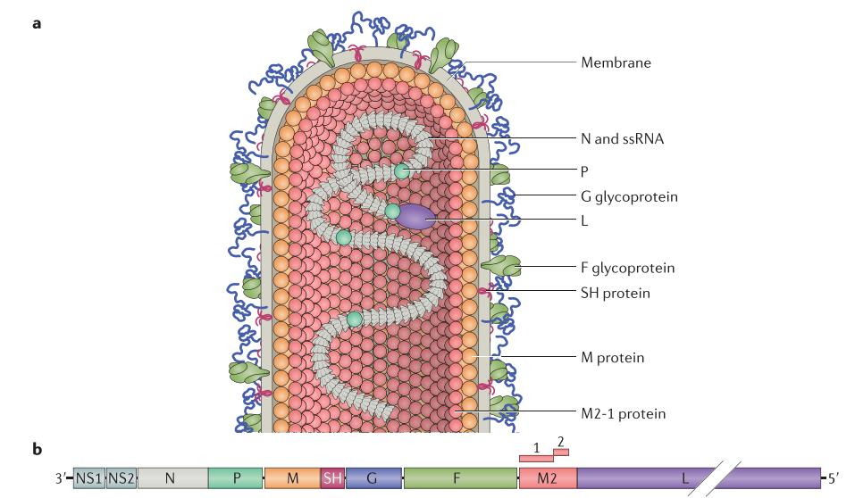 Fig. 1 Respiratory syncytial virus virion
G and F are the major glycoproteins on the surface of the virion and have important roles in entry. The G glycoprotein functions primarily as an attachment protein that binds virions to target cells by interacting with one or more host cell surface molecules. The F glycoprotein can also facilitate attachment, although to a lesser extent than G, but its primary function is to mediate fusion of the viral and host cell membranes[2, 3].
There have been many candidate cellular receptors described for RSV entry, including annexin II, CX3 chemokine receptor 1 (CX3CR1), epidermal growth factor (EGF) receptor, calcium-dependent lectins, Toll-like receptor 4 (TLR4), intercellular adhesion molecule 1 (ICAM-1), nucleolin, and heparan sulfate proteoglycans (HSPGs). Some receptors like EGF are purportedly used by only certain strains of RSV[1].
The RSV-G glycoprotein binds HSPGs, which are abundant on numerous cell types and especially so on many immortalized cell lines, including HEp-2 cells[4]. RSV-G also binds to the CX3C chemokine receptor 1 (CX3CR1) expressed on the apical surface of ciliated bronchial epithelial cells[5]. Although the RSV-G glycoprotein is dispensable for infection, many initial studies characterizing the protein were done using cell lines that express high levels of HSPGs (which also bind RSV-F). When tested in vivo, mice deficient for CX3CR1 were significantly less susceptible to RSV infection. Additionally, the interaction between RSV-G and CX3CR1 induces cellular signaling because it is capable of mediating leukocyte chemotaxis[1, 5].
Nucleolin as a receptor for RSV binds to RSV-F[6]. Expression of human nucleolin on insect cells that are not normally infectible by RSV made them susceptible to infection. Although nucleolin is a predominantly nucleolar protein, a small fraction can be found on the cell surface in vitro and in vivo. This cell surface nucleolin has been implicated as a receptor for a number of viral and bacterial pathogens along with various growth factors.
In addition to CX3CR1 and nucleolin, RSV-F and -G glycoproteins bind to a number of other receptors during cellular entry, shown as in Fig 2.
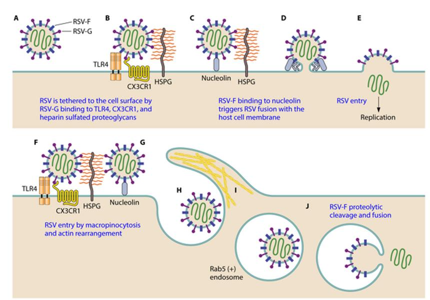 Fig 2 Binding and entry of RSV into the host cell.
The infectious cycle of respiratory syncytial virus (RSV) begins upon attachment of the virion to the apical surface of polarized, ciliated airway epithelial cells (Fig 3). (1) The virion initially binds to the host cell through its G protein and membrane fusion is mediated by the F protein, which anchors into the membrane of the target cell and then folds on itself to bring the viral and host membranes into contact, resulting in membrane fusion. (2) The genome of the virus is used for protein synthesis, with large amounts of NS1/2 and sG protein produced shortly after infection. These proteins protect the replicating virus from the host immune defences. (3) The viral genome is replicated and structural proteins are produced. Transcription and replication occur in the cytoplasm in viral inclusion bodies that serve to concentrate viral products. The viral RNA-dependent RNA polymerase (RdRp) complex is responsible for transcribing viral mRNA and synthesizing positive-sense anti-genome intermediates needed for replication of new negative-sense genomes for packaging into virions. In addition to performing non-proofreading polymerase functions, the RdRp caps and polyadenylates viral mRNAs. (4) The surface glycoproteins are synthesised in the Golgi body and deposited in the host membrane. (5) Assembly of the new virion takes place in the cytoplasm, before budding through the host cell membrane, picking up its surface glycoproteins as part of this process. sG protein is also released. Assembly of RSV virions occurs at or near the plasma membrane. Initial models posited that F proteins associate with lipid rafts and, through the F cytoplasmic tail, recruit and concentrate matrix (M) proteins; this process initiates filament budding through actin-dependent outward membrane deformation[2].
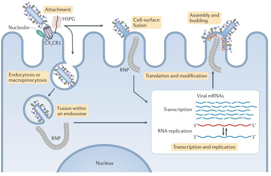 Fig 3 Binding and entry of RSV into the host cell.
Extensive review of RSV replication, pathogenesis, and immune response in animal models and human infections has identified several key issues for RSV vaccine development. First, the immune system of young infants, the primary population for vaccination, is notable for its immaturity and the presence of maternal anti-RSV antibodies during the first several months of life; in turn, these two factors may negatively affect the emergence of a robust immune response following vaccination. Second, a successful RSV vaccine must be able to target both A and B subtypes; this requirement is hampered by the genetic variability of RSV genome, and particularly of the RSV G protein, as well as the post-translational glycosylation of F and G proteins. Third, immunity against RSV remains incomplete after natural infection and thus annual vaccinations may be required; the goal of such vaccination programs is to prevent severe lower respiratory tract infections. It is possible that two vaccines--one for the RSV-naïve, infant population and another for RSV-experienced adults--may be required. Lastly, in the historical context of formalin-inactivated RSV vaccine trials, the safety profile of RSV vaccine candidates will need to be well established during preclinical and clinical development[7].
Various strategies have been pursued to develop an effective and safe RSV vaccine including: 1) inactivated virus preparations; 2) live attenuated/genetically engineered viruses; 3) purified RSV protein subunit vaccine preparations; 4) vector-based vaccine candidates; and 4) DNA-based vaccines. Each approach is summarized below[7].
AREXVY was approved by the US Food and Drug Administration (FDA) on May 3rd, 2023, for the prevention of lower respiratory tract disease (LRTD) caused by respiratory syncytial virus (RSV) in adults aged 60 and older. AREXVY, contains recombinant respiratory syncytial virus glycoprotein F stabilized in the prefusion conformation (RSVPreF3). In clinical trials, the vaccine was generally well tolerated. The most frequently observed solicited adverse events were injection site pain, fatigue, myalgia, headache, and arthralgia. These were generally mild to moderate and transient. The main clinical study of Arexvy was designed to assess the safety and effectiveness of a single dose administered to individuals 60 years of age and older[8].
ABRYSVO is a respiratory syncytial virus (RSV) prefusion F (RSVpreF) vaccine, to help protect both infants through maternal immunization and older adults. ABRYSVO is indicated for: 1) Passive protection against lower respiratory tract disease (LRTD) caused by RSV in infants from birth through six months of age following maternal immunization during pregnancy. 2) Active immunization of individuals 60 years of age and older for the prevention of LRTD caused by RSV. Two additional clinical trials evaluating ABRYSVO has been initiated . One trial is being conducted in children at higher risk for RSV disease ages two to less than 18 years. A second trial is evaluating adults ages 18 to 60 years at higher risk for RSV due to underlying medical conditions such as asthma, diabetes and COPD, and adults ages 18 and older who are immunocompromised and at high-risk for RSV to further describe the safety of the vaccine[9].
References
| Antibody pairs | |||
| Cat. No. | Product Name | Host Species | Antibody pairs |
| DMABP-L43 | Anti-RSV Mab, clone K469 | Mouse | DMABP-L44 (Cap) |
| DMABP-L44 | Anti-RSV Mab, clone K470 | Mouse | DMABP-L43 (Det) |
| DCABY-4812 | Anti-RSV Mab, Clone N04042232 | Mouse | DCABY-4810 (Cap) |
| DCABY-4811 | Anti-RSV Mab, Clone N06002432 | Mouse | DCABY-4810 (Cap) |
| DCABY-4810 | Anti-RSV Mab, Clone N2232903 | Mouse | self pair, DCABY-4811 (Det), DCABY-4812 (Det) |
| CABT-B335 | Anti-RSV Mab, clone 2312 | Mouse | self pair |
| CABT-CS270 | Anti-RSV F Protein Mab, Clone 413 | Rabbit | CABT-CS269 (Cap) |
| CABT-CS269 | Anti-RSV F Protein Mab, Clone S449 | Rabbit | CABT-CS270 (Det) |
| Biosimilar Antibodies | |||
| Cat. No. | Product Name | Host Species | Application |
| CABT-CS600 | Anti RSV Fusion protein Mab (Palivizumab) | Human | ELISA |
| CABT-Z668H | Anti-RSV Mab, clone RSHZ19 (Felvizumab) | Human | ELISA |
| CABT-NS1224 | Anti-RSV Mab, clone RSHZ19 (Felvizumab) | Ferret | ELISA, FuncS |
| CABT-NS1225 | Anti-RSV Mab, clone RSHZ19 (Felvizumab) | Mouse | ELISA, FuncS |
| Neutralizing/Other antibodies | |||
| Cat. No. | Product Name | Host Species | Application |
| CABT-L6335 | Anti-RSV Mab, clone D25 | Human | Neut |
| CABT-NS1787 | Anti-RSV F Protein Mab, clone 10 | Rabbit | ELISA |
| CABT-L2695 | Anti-RSV Pre-F Protein Mab, clone STC54 | Human | Neut, IF |
| CABT-L2696 | Anti-RSV Pre-F Protein Mab, clone STC54 | Rabbit | Neut, IF |
| CABT-L2689 | Anti-RSV Pre & Post-F Protein Mab, clone STC26 | Human | Neut, IF |
| CABT-L2690 | Anti-RSV Pre & Post-F Protein Mab, clone STC26 | Rabbit | Neut, IF |
| DMABT-Z60929 | Anti-RSV G Protein Mab, clone USX245 | Mouse | ELISA, WB, ICC/IF, IHC-Fr |
| CABT-NS1221 | Anti-RSV G Protein Mab, clone 131-2G | Mouse | ELISA |
| CABT-NS1220 | Anti-RSV G Protein Mab, clone 131-2G | Rabbit | ELISA |
| CABT-NS1222 | Anti-RSV G Protein Mab, clone 131-2G | Human | ELISA |
| DCABY-562 | Anti-RSV NP Mab, clone STW4243 (C034) | Mouse | IHC, ELISA, IF |
| DMABT-H21833 | Anti-RSV NP Mab, clone 240-23I | Mouse | ELISA, IF |
| DCABY-814 | Anti-RSV F Protein Mab, clone 792 | Mouse | ELISA, IF |
| DCABY-561 | Anti-RSV F Protein Mab, clone STW4327 (C027) | Mouse | ELISA, EM, FC, IF, IP, WB |
| DMABT-Z60928 | Anti-RSV Phosphoprotein Mab, clone USXJ204 | Mouse | ELISA, WB, IP, ICC/IF, IHC-Fr |
| DMABT-H21834 | Anti-RSV (type A, B) F Protein Mab, clone 240-9G | Mouse | IF |
| DPATB-H83579 | Anti-RSV Pab | Rabbit | WB |
| DPBT-66923GR | Anti-RSV Pab | Goat | IHC, ELISA, IF |
| DPAB-DC4698 | Anti-RSV F Protein Pab | Rabbit | WB |
| DPAB-DC4696 | Anti-RSV M2 Pab | Rabbit | WB |
| Cat. No. | Product Name | Host | Application |
| DAGC455 | Native RSV Type A Antigen | N/A | N/A |
| DAG3087 | Native RSV (Strain Long) Antigen | Hep-2 cells | ELISA |
| DAG-LL001 | Native RSV Type B Lysate | Vero cells | WB, immunoassays |
| DAG-WT315 | RSV F Protein [His] | Mammalian Cell | ELISA, Lateral Flow |
| DAG-H10339 | RSV F Protein (Met 1-Asn 524) [His] | Insect Cells | N/A |
| DAG-H10334 | RSV F Protein (A2, Met 1-Thr 529) [His] | Insect Cells | N/A |
| DAG-H10340 | RSV F Protein (RSS-2, Met 1-Thr 529) [His] | Insect Cells | N/A |
| DAG-WT1180 | RSV Post-F Protein F0 | Mammalian Cell | Immunogen, calibrator, standard |
| DAG-WT1165 | RSV Pre-F Protein F0 | Mammalian Cell | Immunogen, calibrator, standard |
| DAG-H10336 | RSV G Protein (B1, His 67-Ala 299) [His] | HEK293 Cells | N/A |
| DAG-H10338 | RSV G Protein (Asn 66-Arg 297) [His] | HEK293 Cells | N/A |
| DAG-H10337 | RSV G Protein (Asn 66-Arg 297) [His] | Insect Cells | N/A |
| DAG2322 | RSV NP (a.a. 1-391) [His] | E.coli | WB, ELISA, Immunogen |
| DAG2607 | RSV Active Src Protein [GST] | Insect Cells | N/A |
| Cat. No. | Product Name | Species Reactivity | Sample type |
| DEIA1602 | RSV IgM ELISA Kit, Qualitative | Human | Serum, citrate plasma |
| DEIA373 | RSV IgG ELISA Kit, Quantitative | Human | Serum, plasma |
| DEIA374 | RSV IgA ELISA Kit, Quantitative | Human | Serum, plasma |
| DEIA375 | RSV IgM ELISA Kit, Quantitative | Human | Serum, plasma |
| DEIA1788 | RSV IgA ELISA Kit, Qualitative | Human | Serum |
| DEIA1789 | RSV IgG ELISA Kit, Semi-quantitative | Human | Serum |
| DEIA1790 | RSV IgM ELISA Kit, Qualitative | Human | Serum |
CD Creative Diagnostics의 모든 제품을 만나보세요!
Products
Self-Assembled Monolayers Reagents
Protein-Based Fluorescent Nanoparticles
CD Creative Diagnostics - Official Distributor in South Korea "Morebio" 한국 공식 대리점 "모아바이오"


 MSDS
MSDS 온라인 견적


