

다양한 분야에서 혁신적인 솔루션을 지원합니다.
High-Risk HPV L1 VLPs Produced in Yeast provide recombinant virus-like particle antigens for HPV-related research, diagnostic assay development, antibody evaluation, and viral antigen characterization. These antigens are based on the major capsid protein L1 from clinically important high-risk human papillomavirus types. When properly expressed and processed, L1 can self-assemble into virus-like particles that resemble the outer architecture of native HPV capsids while lacking viral genomic material. This makes L1 VLPs non-infectious antigenic reagents that are particularly useful for studies requiring native-like capsid presentation.
The product portfolio covers HPV16, HPV18, HPV31, HPV33, HPV35, HPV45, HPV52, HPV59, and HPV68. This broad panel enables side-by-side comparison of type-specific antibody responses, cross-reactivity, and antigen performance across multiple high-risk genotypes. Yeast-based production supports scalable recombinant expression and provides a practical platform for preparing consistent antigen lots for serology, immunoassay development, and vaccine-related research workflows.
Human papillomaviruses are non-enveloped DNA viruses with capsids primarily composed of L1 and L2 structural proteins. L1 is the principal capsid protein and the major determinant of VLP assembly. Recombinant L1 proteins from many HPV types can assemble into particles that mimic the morphology and surface epitope display of native virions. Because these VLPs do not contain viral DNA, they cannot replicate or initiate infection, but they retain important conformational epitopes for antibody binding.
For HPV serology and antibody studies, conformational integrity is critical. Linear peptides or denatured L1 proteins may fail to represent neutralizing or type-specific epitopes that depend on the three-dimensional arrangement of L1 capsomers. In contrast, properly assembled L1 VLPs present repetitive, native-like surface structures that are well suited for ELISA, bead-based multiplex assays, antibody screening, immunogenicity assessment, and comparative antigenicity studies.
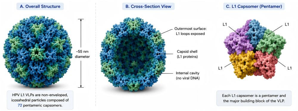 Fig. 1. HPV conformation
HPV infection begins when micro-abrasions expose basal epithelial cells to viral particles. The virion first attaches to heparan sulfate proteoglycans on the basement membrane or cell surface. This initial interaction triggers conformational changes in the capsid, involving exposure and processing of the minor capsid protein L2, and promotes subsequent engagement with entry-associated host factors. The virus is then internalized mainly through endocytic pathways and trafficked through intracellular compartments.
After entry, the viral genome is transported toward the nucleus. Uncoating releases the circular double-stranded DNA genome, which is maintained as an episome in infected basal cells. Early viral proteins, including E1 and E2, regulate viral DNA replication and transcription. High-risk HPV oncogenes E6 and E7 are especially important in disease mechanism research because they can interfere with host tumor suppressor pathways and promote cell cycle progression. Persistent expression of E6 and E7 is a key molecular feature associated with HPV-driven carcinogenesis.
The HPV life cycle is tightly linked to epithelial differentiation. In basal and parabasal layers, the virus maintains its genome at low copy number. As infected cells migrate upward and differentiate, viral genome amplification increases and late structural proteins L1 and L2 are expressed. Assembly of progeny virions occurs in differentiated epithelial cells, followed by release from the epithelial surface. This differentiation-dependent cycle explains why HPV can persist in epithelial tissue and why capsid antigens such as L1 are central to immune recognition, serological assay development, and VLP-based vaccine research.
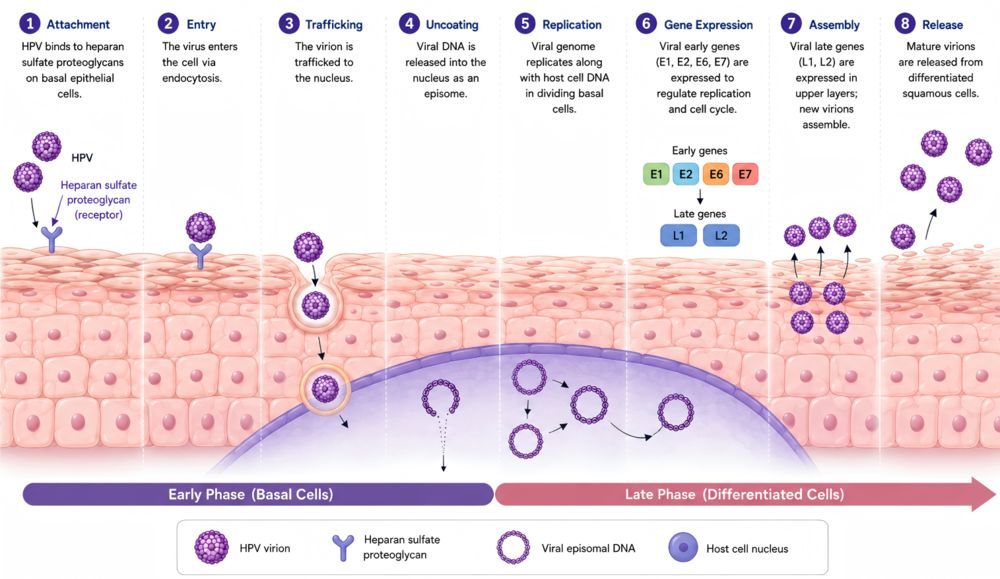 Fig. 2. HPV infection mechanism
| Cat. No. | Product Name | Expression System |
| DAG-WT7707 | Recombinant HPV type 16 L1 VLP | Yeast |
| DAG-WT7708 | Recombinant HPV type 18 L1 VLP | Yeast |
| DAG-WT7709 | Recombinant HPV type 31 L1 VLP | Yeast |
| DAG-WT7710 | Recombinant HPV type 33 L1 VLP | Yeast |
| DAG-WT7711 | Recombinant HPV type 35 L1 VLP | Yeast |
| DAG-WT7712 | Recombinant HPV type 45 L1 VLP | Yeast |
| DAG-WT7713 | Recombinant HPV type 52 L1 VLP | Yeast |
| DAG-WT7714 | Recombinant HPV type 59 L1 VLP | Yeast |
| DAG-WT7715 | Recombinant HPV type 68 L1 VLP | Yeast |
Yeast expression systems provide an efficient and scalable route for producing recombinant HPV L1 antigens. They support controlled expression workflows, practical scale-up, and reproducible production of type-specific L1 VLPs for research and assay development. For laboratories that require multiple HPV genotypes under comparable testing conditions, a yeast-expressed panel offers a consistent source of recombinant antigen material.
The VLP format is the central feature of recombinant HPV L1 antigens. L1 VLPs resemble native capsids in morphology and surface antigen display, but they lack viral DNA and are non-infectious. Their native-like particle structure helps preserve conformational epitopes that are often required for meaningful antibody recognition and serological assay performance.
The portfolio includes nine high-risk HPV types relevant to cervical cancer research, HPV epidemiology, and immune response profiling. Because HPV antibody responses are often type-specific, access to multiple L1 VLPs supports cross-reactivity evaluation, comparative antigen testing, and development of broader serological panels.
Reliable immunoassay development depends on consistent antigen quality, including purity, particle assembly, antigenicity, and coating behavior. Recombinant VLP antigens prepared through controlled production and quality assessment can help reduce assay variability and support more reproducible optimization of ELISA, multiplex, and antibod
Yeast-expressed HPV L1 VLPs may be used as coating or capture antigens in ELISA, chemiluminescent immunoassays, bead-based multiplex assays, and other antibody detection formats. Their conformational epitope display is useful when assay performance depends on recognition of native-like capsid surfaces.
The VLP panel supports evaluation of monoclonal antibodies, polyclonal sera, immune sera, and antibody libraries. Researchers can compare binding across HPV types to assess type specificity, cross-reactivity, and antigenic differences among high-risk genotypes.
Licensed HPV vaccines are based on L1 VLP technology, and recombinant L1 VLPs remain important tools for vaccine-related research. These products are intended as research reagents, not finished vaccine products, and can be used to study antigenicity, antibody binding, and immune response profiles associated with HPV VLP antigens.
Multi-type L1 VLP panels can support seroepidemiological studies by enabling detection and comparison of antibodies against multiple high-risk HPV types. This is useful for studying prior exposure, vaccine-associated antibody profiles, and genotype-specific immune patterns in research settings.
For HPV L1 VLP antigens, quality assessment should address both protein composition and particle-related properties. Purity alone is not sufficient; antigen performance also depends on correct assembly and preservation of conformational epitopes.
| Parameter | Recommended Evaluation Method | Purpose |
| Identity | Mass spectrometry, Western blot, sequence confirmation | Confirms HPV type and L1 protein identity |
| Purity | SDS-PAGE, HPLC, densitometry | Evaluates protein purity and contaminant profile |
| Particle formation | Electron microscopy, dynamic light scattering, size-exclusion chromatography | Confirms VLP assembly and particle size distribution |
| Antigenicity | ELISA or antibody-binding assay | Confirms recognition by HPV type-specific antibodies |
| Endotoxin | LAL assay | Supports use in sensitive immunological workflows |
| Lot consistency | Comparative binding assay or SDS-PAGE profile | Evaluates reproducibility across batches |
| Storage stability | Accelerated and real-time stability studies | Assesses antigen performance over time |
CD Creative Diagnostics의 모든 제품을 만나보세요!
Products
Self-Assembled Monolayers Reagents
Protein-Based Fluorescent Nanoparticles
CD Creative Diagnostics - Official Distributor in South Korea "Morebio" 한국 공식 대리점 "모아바이오"


 MSDS
MSDS 온라인 견적


