


Signosis는 생명 과학 연구 및 생물학적 데이터 분석을 위한 혁신적인 솔루션을 제공하는 글로벌 생명과학 기업입니다.
제품 설명
Mouse Eight-ANA ELISA Screen Kit
제품 번호
EA-5102
제품 특징
Introduction
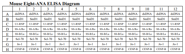
Anti-nuclear antibodies (ANA) are a group of antibodies directed against various nuclear and some cytoplasmic antigens. Serological tests for ANA play an important role towards the diagnosis of various autoimmune connective tissue disorders. Although these antibodies were first associated with systemic lupus erythematosus (SLE), the list of implicated diseases has expanded and many rheumatic diseases are characterized by the presence of one or more of these ANAs. For instance, anti-SSA/Ro and anti-SSB/La antibodies are associated with SLE and Sjogren's Syndrome (SS), anti-dsDNA and anti-Sm antibodies with SLE, antiRNP antibodies with mixed connective tissue disease (MCTD) and SLE, anti-Scl-70 antibodies with scleroderma (progressive systemic sclerosis (PSSJ), anti-Jo1 with polymyositis and dermatomyositis and anti-centromere antibodies with CREST syndrome. As ANA ELISA test collectively detects, in one well, total ANAs against double stranded DNA (dsDNA), Sm, U1-RNP (68K), SS-A/Ro, SSB/La, Scl-70, Jo-1, and centromeric antigens, along with sera positive for IFA HEp-2 ANAs, it is not specific indicators of a connective tissue disease. To monitor more specific antibodies, eight different antigens (dsDNA, SmD1, U1-RNP (68K), SS-A/Ro, SS-B/La, Scl-70, Jo-1, and CENP-B) are coated to different wells in a column or strip for the ELISA screen test of eight different autoimmune antibodies once.
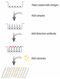
Principle of the assay
In eight-ANA ELISA screen test, eight different antigens (dsDNA, SmD1, U1-RNP (68K), SS-A/Ro, SS-B/La, Scl70, Jo-1, and CENP-B) are coated into different wells in a column or strip. In each well of the strip, one specific antigen is coated for monitoring a corresponding antibody and therefore total 8 wells of a strip allow measurement of 8 different antibodies. The test sample is allowed to react simultaneously with the two components, resulting in antinuclear antibodies being sandwiched between the solid phase and enzyme-linked antibodies. After incubation, the wells are washed to remove unbound-labeled antibodies. A HRP substrate, TMB, is added to result in the development of a blue color. The color development is then stopped with the addition of Stop Solution changing the color to yellow. The concentration of anti-nuclear antibodies is directly proportional to the color intensity of the test sample. Absorbance is measured spectrophotometrically at 450 nm.
Materials provided with the kit
• 8x12 96-well plate coated with 8 different antigens (4oC).
• Anti-mouse IgG antibody conjugated to HRP (4oC).
• 1X Diluent buffer (4oC).
• 5X Assay wash buffer (4oC).
• Substrate (4oC).
• Stop Solution (4oC) Material required but not provided
• Microplate reader capable of measuring absorbance at 450 nm
• Shake
Reagent preparation before starting experiment
• Dilute the 5x Assay wash buffer to 1x buffer 40ml 5x Assay wash buffer 160ml ddH2O
• Dilute 1000 times of anti-mouse IgG antibody conjugated to HRP with 1X Diluent buffer.
Storage and Preparation Store all reagents at 2-8°C.
All reagents must be brought to room temperature (20- 25°C) prior to use.
When stored at 2-8°C, the diluted Assay wash buffer is stable until the kit expiration date.
Assay procedure
1. Calculate the number of samples to decide how many strips need to be used.
2. Add 100μl of Diluent buffer to the wells to be used. Then add 1μl of sample directly in the well to make a 1:100 dilution. Incubate for 1 hour at room temperature with gentle shaking. *Note: We recommend having a blank condition. For the blank, add only diluent buffer to the well.
3. Aspirate each well and wash by adding 200µl of 1X Assay wash buffer. Repeat the process twice for a total of three washes. Completely remove liquid at each wash by firmly tapping the plate against clean paper towels.
4. Add 100µl of diluted anti-mouse IgG antibody conjugated to HRP to each well and incubate for 30 minutes at room temperature with gentle shaking.
5. Repeat the aspiration/wash as in step 3.
6. Add 100µl of Substrate to each well and incubate for 10- 30 minutes.
7. Add 50µl of Stop solution to each well. The color in the wells should change from blue to yellow.
8. Determine the optical density of each well with a microplate reader at 450 nm within 30 minutes.
Signosis의 모든 제품을 만나 보세요!
Stable Cell Lines
Transcription Factor Reporter Cell Lines
HISPEC Nuclear Receptor Reporter Cells
EGFR/HER2/FLT3 Mutant Stable Cell Lines
Stable Cell Line Related Products
TF Assays
TF Luciferase Reporter Vectors
Associate Kit and Key Components
Cytokine ELISA
Associated Kits and Components Cytokine
Gene Expression & miRNA
Key Components: miRNA Northern Blot
miRNA Luciferase Reporter Vectors
Biomarkers
Mitochondria Activity Assay Kit
Oxidative Stress Detection Assay Kits
Signosis - Exclusive Distributor in South Korea "Morebio" 한국 독점 대리점 "모아바이오"


