

Signosis는 Reporter Stable Cell Lines 제품군과 연관 키트들을 전문적으로 제공하는 미국의 제조업체 입니다.
제품 설명
AP-1 Luciferase Reporter HeLa Stable Cell Line (2 vials)
제품 번호
SL-0019
제품 특징
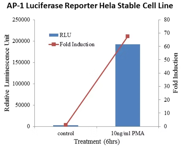
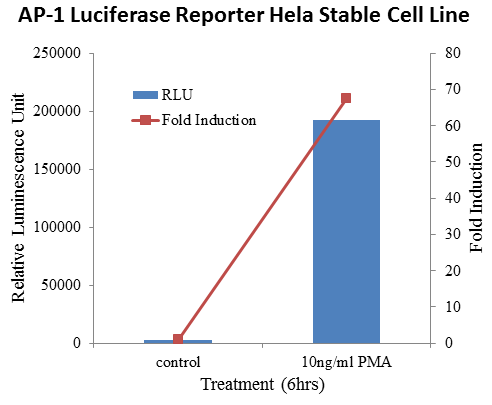
AP-1 Responsive Luciferase Reporter HeLa Stable Cell Line is derived from human cervical cancer, and stably express firefly luciferase reporter gene under the control of AP-1 response element. This cell line is an ideal cellular model for monitoring the activation of JNK, ERK, MAPK Signaling Receptor Signaling Pathway triggered by stimuli treatment, enforced gene expression and gene knockdown.
AP-1 plays a key role in the control of cell proliferation, differentiation, transformation, survival and apoptosis. cJun and Fos dimerize to form AP-1 and this protein complex selectively binds to DNA sequences containing the TPA-responsive elements (TREs) to regulate gene expression. Many stimuli, both physiological and pathological, can regulate AP-1 activity, including TPA, serum, growth factors, cytokines, stress signals, infections, and oncoproteins, TNF and IL-1. AP-1 activity can be used as readout for JNK, ERK, or MAPK kinase signaling pathway.
Signosis has developed AP-1 luciferase reporter HeLa stable cell line by transducing cells with baculovirus containing both AP-1 luciferase reporter and hygromycin expression cassette. The hygromycin resistant clones were subsequently screened for PMA-induced luciferase activity. The cell line can be used as a reporter system for monitoring the activity of AP-1 triggered by stimuli treatment, such as TNFa, IL-1, gene overexpression and gene knockdown. The cells contain no viral particles and require handling at biosafety level 1 protocol.
Principle behind TF luciferase reporter. TF luciferase reporter stable cell line utilizes artificial promoter constructs to drive luciferase expression. The promoter region can consists of multiple repeats of a cis-element TF binding site, a DNA fragment from the promoter region of a known TF downstream gene, or a DNA fragment containing putative/known TF binding sites. There are several ways that a TF can be activated, such as through extracellular stimuli or through intracellular signaling pathways. Once activated, the TF translocates to the nucleus and often interacts with relevant co-factors to drive gene expression. Once luciferase is expressed, it can generate light in an enzymatic assay and the amount of light measured is positively correlated with the level of TF activation.
Signosis의 모든 제품을 만나보세요!
Stable Cell Lines
Transcription Factor Reporter Cell Lines
HISPEC Nuclear Receptor Reporter Cells
EGFR/HER2/FLT3 Mutant Stable Cell Lines
Stable Cell Line Related Products
TF Assays
TF Luciferase Reporter Vectors
Associate Kit and Key Components
Cytokine ELISA
Associated Kits and Components Cytokine
Gene Expression & miRNA
Key Components: miRNA Northern Blot
miRNA Luciferase Reporter Vectors
Biomarkers
Signosis - Exclusive Distributor in South Korea "Morebio" 한국 독점 대리점 "모아바이오"


 MSDS
MSDS 온라인 견적