


YEASEN은 생명과학 연구 및 진단을 위한 고품질의 연구용 시약과 기기를 제공하는 글로벌 바이오 기업입니다.
제품 설명
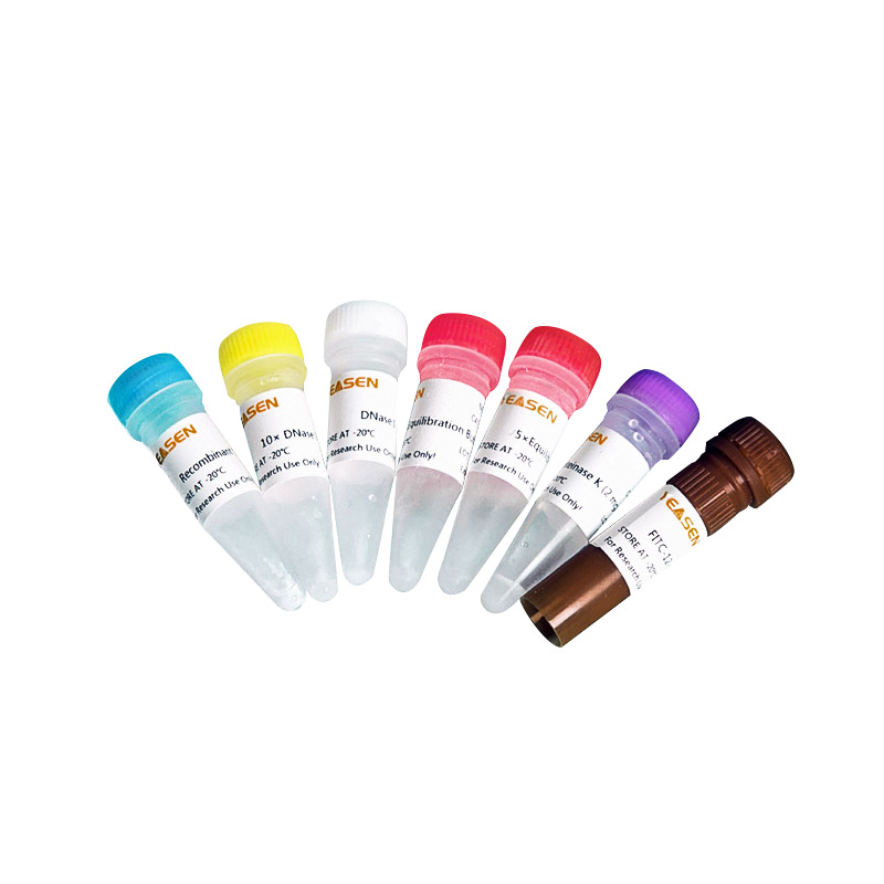
TUNEL Apoptosis Detection Kit (FITC)
제품 번호
40306ES20 / size: 20T
40306ES50 / size: 50T
40306ES60 / size: 100T
제품 설명
Product name | Cat# | Specification |
| 40306ES20 | 20 T |
TUNEL Apoptosis Detection Kit (FITC) TUNEL | 40306ES50 | 50 T |
| 40306ES60 | 100 T |
When cells undergo apoptosis, they activate endonuclease enzymes that cut genomic DNA between nucleosomes. DNA ladder of 180-200 bp can be detected by electrophoresis after extraction of DNA during apoptosis.
TUNEL (TdT mediated dUTP Nick End Labeling) Cell apoptosis Assay Kit (FITC) can be used to detect the nuclear DNA breakage in tissue cells during late apoptotic stage. The principle is that FITC-12-dUTP is inserted into the 3´-hydroxyl (3´-OH) terminal of genomic DNA exposed during DNA break under the action of Terminal Deoxynucleotidyl Transferase (TdT). This can be detected by fluorescence microscopy or flow cytometry.
This kit optimizes the labeling reaction by using FITC-12-dUTP and unlabeled dNTP at the optimal ratio for nucleotide incorporation at the 3 '-OH terminal, resulting in a longer "labeling tail" at the end of the same broken DNA fragment. The "labeling tail" reduces the steric hindrance of labeled groups on adjacent dNTP, increases the number of fluorescent-groups on each broken fragment, and reduces the aggregation and quenching that may be caused by adjacent fluorescent-groups, thus improving detection sensitivity and reducing non-specific reactions.
This kit has a wide range of applications. It can be used to detect apoptosis in frozen or paraffin sections, as well as in culture adherent cells or suspension cells situation.
The components are shipped with ice pack and can be stored at -20°C for 1 year.
A. Paraffin-embedded tissue section
1. Soak paraffin sections in xylene for 5 mins at room temperature, repeat once to remove paraffin thoroughly.
2. Soak the slices with 100% ethanol at room temperature for 5 mins, repeat once.
3. Soak with gradient ethanol (90, 80, 70%) for 3 mins at room temperature.
4. Wash sections gently with PBS and carefully blot the excess liquid around the sample on the slide with filter paper.
5. Preparation of Proteinase K working solution: Dilute 2 mg/mL Proteinase K solution with PBS in the ratio of 1:100, so that the final concentration is 20 μg/mL.
6. Add 100 μL of Proteinase K working solution to each sample, cover it completely, and incubate for 20 mins at room temperature.
【Notes】: Proteinase K helps tissues and cells to permeate the staining reagent in subsequent steps. Too long incubation time will increase the risk of tissue slices falling off from carrier slices in subsequent washing steps, while too short incubation time may cause inadequate permeability treatment and affect labeling efficiency. Without better results, it may be necessary to optimize the incubation time of Proteinase K.
7. Rinse the sample with PBS solution, gently remove the excess liquid, and carefully blot the liquid around the sample on the slide with filter paper. The treated samples are placed in a wet box to keep them moist.
B. Frozen tissue section
1. The slides were immersed in 4% paraformaldehyde solution (dissolved in PBS) and fixed, and incubated at room temperature for 15 mins.
2. Gently remove excess liquid and carefully blot the excess liquid around the sample on the slide with filter paper.
3. The slides were immersed in PBS solution and incubated at room temperature for 15 mins.
4. Gently remove excess liquid and carefully blot the excess liquid around the sample on the slide with filter paper.
5. Preparation of Proteinase K working solution: Dilute 2 mg/mL Proteinase K solution with PBS in the ratio of 1:100, so that the final concentration is 20 μg/mL.
6. Add 100 μL of Proteinase K working solution to each sample, cover it completely, and incubate at room temperature for 10 mins.
【Notes】: Proteinase K helps tissues and cells to permeate the staining reagent in subsequent steps. Too long incubation time will increase the risk of tissue slices falling off from carrier slices in subsequent washing steps, while too short incubation time may cause inadequate permeability treatment and affect labeling efficiency. Without better results, it may be necessary to optimize the incubation time of Proteinase K.
7. Rinse samples 2-3 times with PBS solution.
8. Gently remove excess liquid and carefully blot the liquid around the sample on the slide with filter paper. The treated samples are placed in a wet box to keep them moist.
C. Cell sample
[Preparation of cell slipper]
Adherent cells were cultured in Lab-Tek Chamber Slides. After apoptosis induction, the slides were washed twice with PBS.
Preparation of cell smear (polylysine-coated slide as an example)
1. Prepare poly-lysine coated slides: Drain 50 - 100 μL 0.01% (w/v) poly-lysine solution onto the surface of each pre-cleaned glass slide. Spread the polylysine solution in a thin layer over the area to be used for cell fixation. After the slides are dried, rinse them quickly with deionized water and let the coated slides dry in air for 30-60 minutes. Coated slides can be stored at room temperature for several months.
2. The cells were re-suspended in PBS at a concentration of about 2×10⁷ cells /mL, and 50-100 μL cell suspension drops were absorbed onto polylysine-coated slides, and the cell suspension was gently coated with a clean slide.
Follow the following steps to process the cell samples:
1. The cells were fixed, and the slides were immersed in a dye vat containing 4% fresh paraformaldehyde prepared in PBS, and placed at 4℃ for 25 mins.
2. Wash the slides, immerse them in PBS, and leave them at room temperature for 5 mins. Wash with PBS again.
3. Gently remove excess liquid and carefully blot the excess liquid around the sample on the slide with filter paper.
4. Each sample can be immersed in 0.2% Triton X-100 solution prepared in PBS and incubated for 5 mins at room temperature for transparent treatment.
5. Immerse and clean the sample 2-3 times in an open beaker containing PBS solution.
6. Gently remove excess liquid and carefully blot the liquid around the sample on the slide with filter paper. The treated samples are placed in a wet box to keep them moist.
2. Steps of DNA enzyme treatment of positive control (optional)
After the permeable treatment of samples, cells were treated with DNA enzyme I to prepare positive control slides. This process usually causes most of the treated cells to glow green.
【Notes】: The treatment of fixed cells by DNA enzyme I can cause the breakage of chromosomal DNA, resulting in many labeled DNA 3 '- ends.
3. Tap the slide to remove excess liquid and wash the slide thoroughly 3-4 times in a dye vat with deionized water.
【Notes】: A separate dyeing cylinder must be used for the positive control slides, otherwise the residual DNase I on the positive control slides may introduce a high background to the experimental slides.
3. Marking and detection
1. Dilute 5× the equilibration buffer with deionized water at a ratio of 1:5.
2. Add 100 μL 1×Equilibration Buffer to each sample to cover the sample area and incubate with Equilibration buffer at room temperature for 10-30 mins. Or place slides into a vat with 1×slides bration buffer to ensure that the buffer does not reach the sample. FITC-12-dUTP Labling Mix was thawed on ice while the cells were balanced, and an adequate amount of TdT incubation buffer was prepared for all experimental and optional positive control reactions as per Table 1. For a standard reaction with an area less than 5 cm2 and a volume of 50 μL, multiply 50 μL by the number of experimental and positive control reactions to determine the total volume of TdT incubation buffer required. For samples with larger surface areas, the reagent volume can be increased proportionally.
Table 1. TdT incubation buffers prepared for experimental and optional positive control reactions
Component | Volume (μL /50 μL system) |
ddH2O | 34 |
5×Equilibration Buffer | 10 |
FITC-12-dUTP Labling Mix | 5 |
Recombinant TdT Enzyme | 1 |
Negative control system: Prepare a control incubation buffer without TdT enzyme and replace TdT enzyme with ddH2O.
3. Wash most of the 100 μL 1×Equilibration Buffer with absorbant paper around the Equilibration area, and add 50 μL TdT to the 5 cm2 area cells incubation buffer. Don't let the cells dry out. After that, the slides are shielded from light.
4. Place a plastic cover glass over the cells to ensure an even distribution of the reagent, and place a paper towel soaked in water on the bottom of the wet box. The slides were placed in a wet box and incubated at 37℃ for 60 mins. Cover the wet box with aluminum foil to protect it from light.
【Notes】: Plastic cover glass can be cut in half before use. Fold the edge of the cover glass for easy removal and manipulation.
5. Remove the plastic cover glass and incubate the sections in PBS solution at room temperature for 5 mins.
6. Gently remove the excess liquid and replace it with fresh PBS solution and incubate for 5 mins at room temperature. Repeat.
7. Gently wipe the PBS solution around and on the back of the sample with filter paper.
【Notes】: In order to reduce the background, the slides can be washed with PBS containing 0.1% Triton X-100 and 5 mg/mL BSA for 3 times, 5 min each, after washing once with PBS, so that the free unreacted markers can be cleaned.
9. Wash the samples, immerse the slides in deionized water, place at room temperature for 5 mins, repeat twice, wash three times in total.
10. Remove excess water off the slide and wipe the area around the cells with absorbent paper.
11. Samples were immediately analyzed under a fluorescence microscope, and green fluorescence was observed under 520±20 nm fluorescence with a standard fluorescence filter; Red fluorescence of PI was observed at > 620 nm, or blue DAPI was observed at 460 nm. Slides can be stored overnight in 4℃ darkness if necessary. PI/DAPI could color both apoptotic and non-apoptotic cells red/blue, and only the apoptotic nuclei had FITC-12-dUTP incorporation and localization of green fluorescence.
4. Flow cytometry was used to detect suspended cells
1. 3-5×10⁶ cells were washed twice by centrifugation at 4℃ (300×g) with PBS, and then re-suspended in 0.5 mL PBS.
2. Fixed cells, added 5 mL 1% paraformaldehyde solution in PBS, and placed on ice for 20 mins.
3. The cells were centrifuged at 4℃ for 10 mins at 300×g, then supernatant was removed and re-suspended in 5mL PBS. The cells were washed repeatedly and resuspended with 0.5 mL PBS.
4. Permeable cells were added with 5 mL 70% ethanol precooled on ice and incubated at -20℃ for 4 hours. Cells can be stored in 70% ethanol at -20℃ for one week, or cells can be permeated in 0.2% Triton X-100 solution prepared in PBS at room temperature for 5 mins.
5. Cells were centrifuged at 300×g for 10 mins and resuspended with 5 mL PBS. Centrifugation was repeated and 1 mL PBS was resuspended.
6. 2×10⁶ cells were transferred to a 1.5 mL microcentrifuge tube.
7.300×g centrifugation for 10 mins, the supernatant was removed, and the slides were re-suspended with 80 μL 1× Slides Buffer. Incubate at room temperature for 5 mins.
8. While balancing the cells, melt the FITC-12-dUTP-labeled mixture on ice and prepare a sufficient amount of TdT incubation buffer for all reactions as per Table 1. For a standard reaction of 2×10⁶ cells with a volume of 50 μL, multiply 50 μL by the number of reactions to determine the total volume of TdT incubation buffer required.
9. The cells were centrifuged at 300× g for 10 mins, the supernatant was removed, and the precipitate was suspended in 50 μL TdT incubation buffer, and incubated at 37℃ for 60 mins, away from light. The cells were gently resuspended with a micropipette every 15 mins.
10. Add 1 mL, 20 mM EDTA to stop the reaction, and gently mix with a micropipette.
11. 300×g centrifugation for 10 mins, supernatant was removed and the precipitate was re-suspended in 1mL 0.1% Triton X-100 solution prepared in PBS, containing 5 mg/mL BSA, and washed twice in total.
14. Cells were analyzed by flow cytometry and measured for FITC-12-dUTP green fluorescence at 520 ± 20 nm and PI red fluorescence at > 620 nm. PI dyed both apoptotic and non-apoptotic cells red, and only FITC-12-dUTP was incorporated into the apoptotic nuclei to locate the green fluorescence.
YEASEN의 모든 제품을 만나 보세요!
YEASEN - Official Distributor in South Korea "Morebio" 한국 공식 대리점 "모아바이오"


