

Signosis는 생명 과학 연구 및 생물학적 데이터 분석을 위한 혁신적인 솔루션을 제공하는 글로벌 생명과학 기업입니다.
제품 설명
Mouse Anti-Scl-70 ELISA Kit
제품 번호
EA-5205
제품 특징
Introduction
Antibodies to Scl-70 are a specific immunological marker for scleroderma (or progressive systemic sclerosis, PSS), a systemic autoimmune disease characterized by collagen deposition and connective tissue destruction of the skin, blood vessels and certain internal organs. Studies have shown varying frequencies of Scl-70 antibodies in PSS. This antibody was found in approximately 20% of PSS patients in early studies but 75% in later studies. Scl-70 antibodies are directed against DNA-topoisomerase I which locates in the nucleus. The whole molecule of DNAtopoisomerase is 110 kDa but it is easily degraded by proteases to 100 kDa, 87 kDa and 70 kDa (Scl-70). PSS is classified into two types; diffuse scleroderma and limited scleroderma. Scl-70 antibodies are present specifically in diffuse scleroderma and centromere antibodies are present in limited scleroderma. Rarely, Scl-70 antibodies are found in SLE and MCTD patients.
Principle of the assay
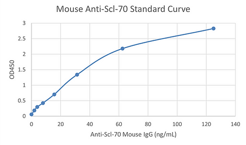
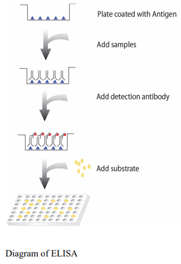
Anti-Scl-70 ELISA kit measures anti-Scl-70 antibodies in the serum. It is based on the principle of a solid phase enzyme-linked immunosorbent assay. The assay utilizes Scl70 protein for immobilization on the microtiter wells and anti-mouse IgG antibodies conjugated to horseradish peroxidase (HRP) for detection. The test sample is allowed to react simultaneously with the two components, resulting in anti-Scl-70 antibodies being sandwiched between the solid phase and enzyme-linked antibodies. After incubation, the wells are washed to remove unbound-labeled antibodies. A HRP substrate, TMB, is added to result in the development of a blue color. The color development is then stopped with the addition of Stop Solution changing the color to yellow. The concentration of anti-Scl-70 is directly proportional to the color intensity of the test sample. Absorbance is measured spectrophotometrically at 450 nm.
Materials provided with the kit
• 8x12 96-well plate coated with Scl-70 (4oC).
• Anti-mouse IgG antibody conjugated to HRP (4oC).
• Mouse Scl-70 Positive Control (4oC).
• 1X Diluent buffer (4oC).
• 5X Assay wash buffer (4 oC).
• Substrate (4oC).
• Stop Solution (4oC).
Material required but not provided
• Microplate reader capable of measuring absorbance at 450 nm
• Shaker
Reagent preparation before starting experiment
• Dilute the 5x Assay wash buffer to 1x buffer 40ml 5x Assay wash buffer 160ml ddH2O
• Dilute 1000 times of anti-mouse IgG antibody conjugated to HRP with 1X Diluent buffer.
Storage and Preparation
Store all reagents at 2-8°C. All reagents must be brought to room temperature (20- 25°C) prior to use.
When stored at 2-8°C, the diluted Assay wash buffer is stable until the kit expiration date.
SAMPLE COLLECTION AND STORAGE
Serum
Use a serum separator tube and allow samples to clot for 30 minutes before centrifugation for 15 minutes at 1000 g. Remove serum and assay immediately or aliquot and store samples at -20° C. Avoid repeated freeze-thaw cycles.
Plasma
Collect plasma using citrate, EDTA, or heparin as an anticoagulant. Centrifuge for 15 minutes at 1000 g within 30 minutes of collection. Assay immediately or aliquot and store samples at -20° C. Avoid repeated freeze-thaw cycles.
Assay procedure
1. Calculate the number of samples to decide how many strips need to be used. Make sure the rest wells are well sealed.
2. Standard Curve if needed
• Add 200 μl 1X Diluent Buffer to the 1st well on one strip
• Add 100 μl 1X Diluent Buffer to the rest of wells on the same strip
• Add appropriate amount of mouse Scl-70 positive control (50 μg/ml) to 1st well as 1st dilution
• Mix 1st dilution in 1st well and transfer 100 μl from 1st to next well for next dilution. Perform six two-fold serial dilutions
• 1X Diluent buffer serves as the zero standard or blank
Note: The first dilution starting from 250 ng/ml is recommended.
3. Add 100 µl of diluted samples or positive control (1:100 diluted with 1X Diluent Buffer) per well and incubate for 1 hour at room temperature with gentle shaking.
*Note: We recommend having a blank condition. For the blank, add only diluent buffer to the well.
3. Aspirate each well and wash by adding 200 µl of 1X Assay wash buffer. Repeat the process twice for a total of three washes. Completely remove liquid at each wash by firmly tapping the plate against clean paper towels. 4. Add 100 µl of diluted anti-mouse IgG antibody conjugated to HRP to each well and incubate for 30 minutes at room temperature with gentle shaking.
5. Repeat the aspiration/wash as in step 3.
6. Add 100 µl of Substrate to each well and incubate for 5-30 minutes.
7. Add 50 µl of Stop solution to each well. The color in the wells should change from blue to yellow.
8. Determine the optical density of each well with a microplate reader at 450 nm within 30 minutes

Signosis의 모든 제품을 만나 보세요!
Stable Cell Lines
Transcription Factor Reporter Cell Lines
HISPEC Nuclear Receptor Reporter Cells
EGFR/HER2/FLT3 Mutant Stable Cell Lines
Stable Cell Line Related Products
TF Assays
TF Luciferase Reporter Vectors
Associate Kit and Key Components
Cytokine ELISA
Associated Kits and Components Cytokine
Gene Expression & miRNA
Key Components: miRNA Northern Blot
miRNA Luciferase Reporter Vectors
Biomarkers
Mitochondria Activity Assay Kit
Oxidative Stress Detection Assay Kits
Signosis - Exclusive Distributor in South Korea "Morebio" 한국 독점 대리점 "모아바이오"


