

Boster Bio는 생명과학 연구를 위한 항체, ELISA 키트, 단백질 및 관련 제품을 제공하는 미국의 생명과학 기업입니다.
제품 설명
Anti-GAA Antibody Picoband® (monoclonal, 2G7)
제품 번호
M01548
제품 특징
| SKU: | M01548 |
|---|---|
| Size: | 100 μg/vial |
| Reactive Species: | Human |
| Host: | Mouse |
| Application: | IF, IHC, ICC, WB |
Anti-GAA Antibody Picoband® (monoclonal, 2G7)
M01548
100 μg/vial
Lyophilized
Boster Bio Anti-GAA Antibody Picoband® (monoclonal, 2G7) catalog # M01548. Tested in IF, IHC, ICC, WB applications. This antibody reacts with Human. The brand Picoband indicates this is a premium antibody that guarantees superior quality, high affinity, and strong signals with minimal background in Western blot applications. Only our best-performing antibodies are designated as Picoband, ensuring unmatched performance.
Store at -20˚C for one year from date of receipt. After reconstitution, at 4˚C for one month. It can also be aliquotted and stored frozen at -20˚C for six months. Avoid repeated freeze-thaw cycles.
Anti-GAA Antibody Picoband® (monoclonal, 2G7) (Boster Biological Technology, Pleasanton CA, USA, Catalog # M01548)
Mouse
Each vial contains 4mg Trehalose, 0.9mg NaCl, 0.2mg Na2HPO4, 0.01mg NaN3.
Monoclonal
2G7
Mouse IgG2b
A synthetic peptide corresponding to a sequence in the middle region of human GAA, different from the related mouse sequence by eight amino acids, and from the related rat sequence by six amino acids.
No cross-reactivity with other proteins.
M01548 is reactive to GAA in Human
110 kDa, 95 kDa, 76 kDa
105.3 kDa
Lysosomal alpha-glucosidase is an enzyme that in humans is encoded by the GAA gene. This gene encodes lysosomal alpha-glucosidase, which is essential for the degradation of glycogen to glucose in lysosomes. The encoded preproprotein is proteolytically processed to generate multiple intermediate forms and the mature form of the enzyme. Defects in this gene are the cause of glycogen storage disease II, also known as Pompe's disease, which is an autosomal recessive disorder with a broad clinical spectrum. Alternative splicing results in multiple transcript variants.
Boster validates all antibodies on WB, IHC, ICC, Immunofluorescence, and ELISA with known positive control and negative samples to ensure specificity and high affinity, including thorough antibody incubations.
M01548 is guaranteed for IF, IHC, ICC, WB Boster Guarantee
| Application | Dilution | Species |
|---|---|---|
| Western blot | 0.1-0.5μg/ml | Human |
| Immunohistochemistry (Paraffin-embedded Section) | 0.5-1μg/ml | Human |
| Immunocytochemistry/Immunofluorescence | 2μg/ml | Human |
Click image to see more details
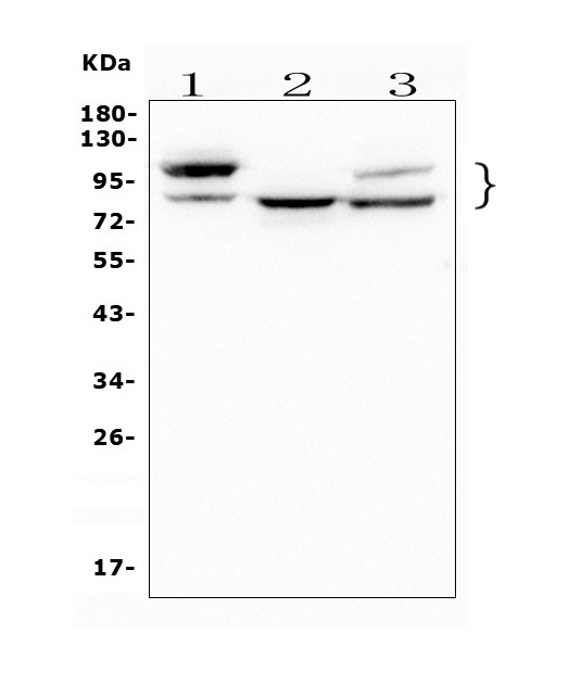
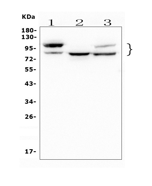
Western blot analysis of GAA using anti-GAAantibody (M01548).
Electrophoresis was performed on a 5-20% SDS-PAGE gel at 70V (Stacking gel) / 90V (Resolving gel) for 2-3 hours. The sample well of each lane was loaded with 50ug of sample under reducing conditions.
Lane 1: human A549 tissue lysates,
Lane 2: human HEK293 whole cell lysates,
Lane 3: human PC-3 whole cell lysates,
After Electrophoresis, proteins were transferred to a Nitrocellulose membrane at 150mA for 50-90 minutes. Blocked the membrane with 5% Non-fat Milk/ TBS for 1.5 hour at RT. The membrane was incubated with mouse anti-GAA antigen affinity purified polyclonal antibody (Catalog # M01548) at 0.5 μg/mL overnight at 4°C, then washed with TBS-0.1%Tween 3 times with 5 minutes each and probed with a goat anti-mouse IgG-HRP secondary antibody at a dilution of 1:10000 for 1.5 hour at RT. The signal is developed using an Enhanced Chemiluminescent detection (ECL) kit (Catalog # EK1001) with Tanon 5200 system. A specific band was detected for GAA at approximately 110,95,76KD. The expected band size for GAA is at 105KD.
Click image to see more details
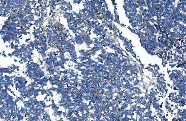
IHC analysis of GAA using anti-GAA antibody (M01548).
GAA was detected in paraffin-embedded section of human lung cancer tissue. Heat mediated antigen retrieval was performed in EDTA buffer (pH8.0, epitope retrieval solution). The tissue section was blocked with 10% goat serum. The tissue section was then incubated with 1μg/ml mouse anti-GAA Antibody (M01548) overnight at 4°C. Biotinylated goat anti-mouse IgG was used as secondary antibody and incubated for 30 minutes at 37°C. The tissue section was developed using Strepavidin-Biotin-Complex (SABC) (Catalog # SA1021) with DAB as the chromogen.
Click image to see more details
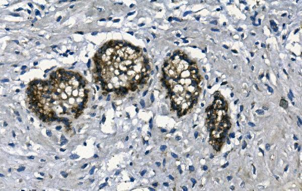
IHC analysis of GAA using anti-GAA antibody (M01548).
GAA was detected in paraffin-embedded section of human mammary cancer tissue. Heat mediated antigen retrieval was performed in EDTA buffer (pH8.0, epitope retrieval solution). The tissue section was blocked with 10% goat serum. The tissue section was then incubated with 1μg/ml mouse anti-GAA Antibody (M01548) overnight at 4°C. Biotinylated goat anti-mouse IgG was used as secondary antibody and incubated for 30 minutes at 37°C. The tissue section was developed using Strepavidin-Biotin-Complex (SABC) (Catalog # SA1021) with DAB as the chromogen.
Click image to see more details
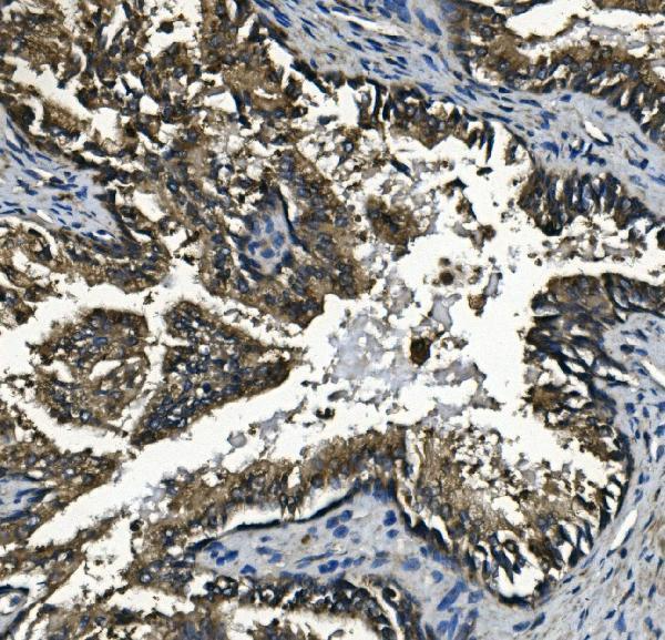
IHC analysis of GAA using anti-GAA antibody (M01548).
GAA was detected in paraffin-embedded section of human prostatic cancer tissue. Heat mediated antigen retrieval was performed in EDTA buffer (pH8.0, epitope retrieval solution). The tissue section was blocked with 10% goat serum. The tissue section was then incubated with 1μg/ml mouse anti-GAA Antibody (M01548) overnight at 4°C. Biotinylated goat anti-mouse IgG was used as secondary antibody and incubated for 30 minutes at 37°C. The tissue section was developed using Strepavidin-Biotin-Complex (SABC) (Catalog # SA1021) with DAB as the chromogen.
Click image to see more details
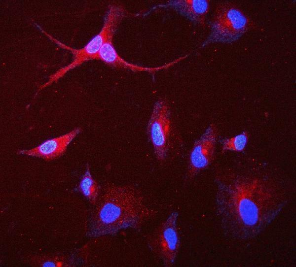
IF analysis of GAA using anti-GAA antibody (M01548).
GAA was detected in immunocytochemical section of A549 cell. Enzyme antigen retrieval was performed using IHC enzyme antigen retrieval reagent (AR0022) for 15 mins. The cells were blocked with 10% goat serum. And then incubated with 2μg/mL mouse anti-GAA Antibody (M01548) overnight at 4°C. Cy3 Conjugated Goat Anti-Mouse IgG (BA1031) was used as secondary antibody at 1:100 dilution and incubated for 30 minutes at 37°C. The section was counterstained with DAPI. Visualize using a fluorescence microscope and filter sets appropriate for the label used.
Click image to see more details
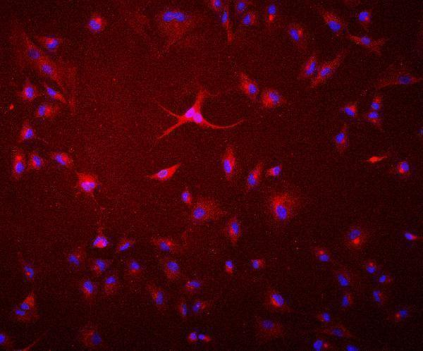
IF analysis of GAA using anti-GAA antibody (M01548).
GAA was detected in immunocytochemical section of A549 cell. Enzyme antigen retrieval was performed using IHC enzyme antigen retrieval reagent (AR0022) for 15 mins. The cells were blocked with 10% goat serum. And then incubated with 2μg/mL mouse anti-GAA Antibody (M01548) overnight at 4°C. Cy3 Conjugated Goat Anti-Mouse IgG (BA1031) was used as secondary antibody at 1:100 dilution and incubated for 30 minutes at 37°C. The section was counterstained with DAPI. Visualize using a fluorescence microscope and filter sets appropriate for the label used.
BosterBio의 모든 제품을 만나 보세요!
BosterBio - Official Distributor in South Korea "Morebio" 한국 공식 대리점 "모아바이오"


 MSDS
MSDS 온라인 견적


